40 label the structures of a skeletal muscle fiber.
Structures of the Skeletal Muscle Fiber Flashcards | Quizlet Muscle Cell or Muscle Fiber or Myofiber -Muscle cells are long, cylindrical & multinucleated -Sarcolemma = muscle cell membrane -Sarcoplasm filled with tiny threads called myofibrils & myoglobin (red-colored, oxygen-binding protein) Transverse Tubules -T (transverse) tubules are invaginations of the sarcolemma into the center of the cell Label structure of skeletal muscle Diagram | Quizlet Label structure of skeletal muscle 4.0 5 Reviews How do you want to study today? Learn Focus your studying with a path Test Take a practice test Match Get faster at matching terms + − Created by danielaaaa04 Terms in this set (8) myofibrils ... sarcoplasmis reticulum ... sarcolemma ... epimysium ... perimysium ... endomysium ... fascicle ...
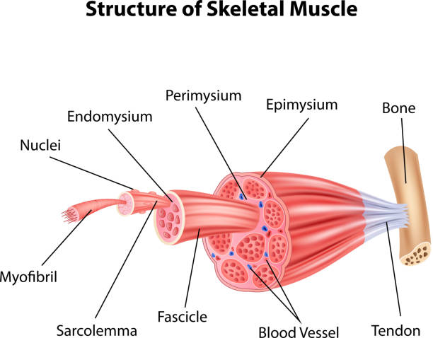
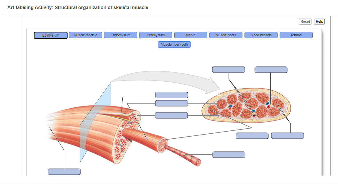
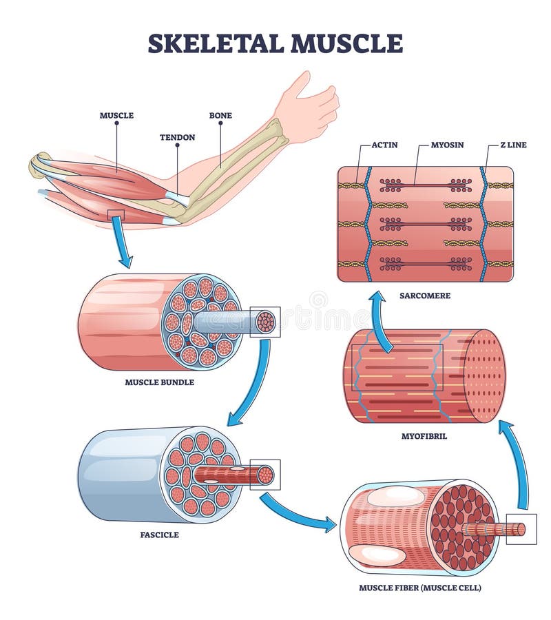
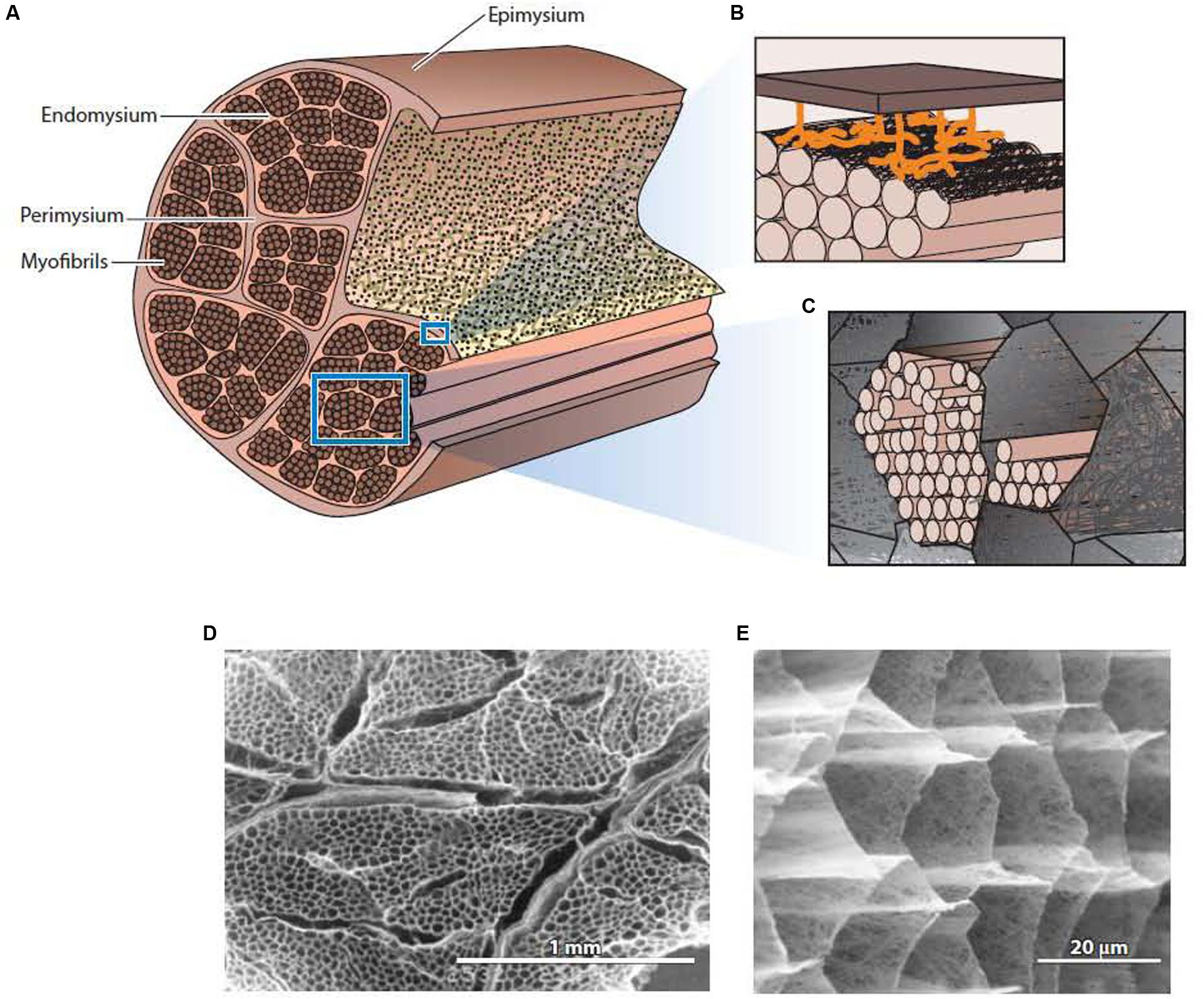
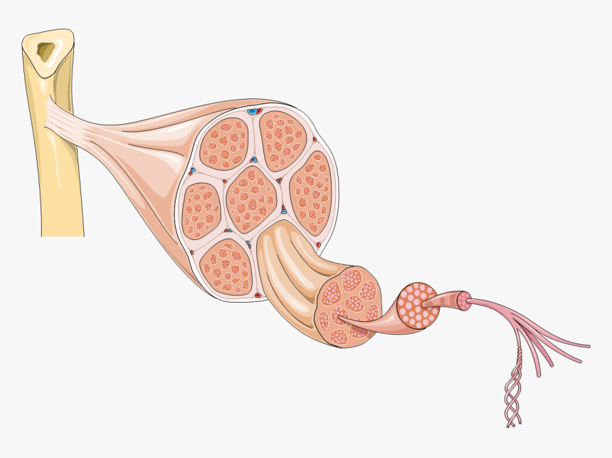
Skeletal Muscle Tissue Anatomy and Structure - Registered Nurse RN Each skeletal muscle is considered an organ, and it's made up of connective tissue layers, muscle fibers, blood vessels, and nerves. Skeletal muscles attach to the bones through tendons or through a direct attachment. As you look at this muscle diagram, you'll notice an outer layer of connective tissue called epimysium.
Label the structures of a skeletal muscle fiber.
Muscle Structure Labeling Quiz - PurposeGames.com This online quiz is called Muscle Structure Labeling. This online quiz is called Muscle Structure Labeling. English en. Login. Login Register Free Help; Start; Explore. Games; Playlists; Tournaments; Tags; The Wall; Badges; ... Skeletal Muscle Matching - Back of the Body 10p Image Quiz. Label Parts of the Skull - Lateral View 10p Image Quiz. Solved Hel Label the structures of a skeletal muscle fiber ... - Chegg Science. Anatomy and Physiology. Anatomy and Physiology questions and answers. Hel Label the structures of a skeletal muscle fiber. 4 0.1 points eBook Sarcoplasmic reticulum Nucleus Myofibril Openings into T tubules Sarcolemma Mc Graw Hill < Prey 4 of 20 !!! 11.2: Microscopic Anatomy of Skeletal Muscles - Biology LibreTexts Microscopic Anatomy of Skeletal Muscles. Skeletal muscle is found attached to bones. It consists of long multinucleate fibers. The fibers run the entire length of the muscle they come from and so are usually too long to have their ends visible when viewed under the microscope. The fibers are relatively wide and very long, but unbranched.
Label the structures of a skeletal muscle fiber.. Solved Muscle Cell Label the structures of a skeletal muscle - Chegg Expert Answer 100% (17 ratings) 1) Sarcolemma 2) myofib … View the full answer Transcribed image text: Muscle Cell Label the structures of a skeletal muscle fiber. Nucleus Myofibril Sarcolemma Sarcoplasmic reticulum Openings into T tubules < Prev 3 of 15 !!! Next > Thinkinys - How to write a boty The Good Cre. Identify the structures of skeletal muscle. - Brainly.com Answer: Skeletal muscle is one of the major muscle types in most animals specialized to perform movement of the human body, maintaining body posture and generation of body heat present in different sizes and shapes. The main structure of skeletal muscle cells consists of various integrated tissues: 1. Muscle: soft, contractile tissue important ... Skeletal muscle tissue: Histology | Kenhub Special terms are used to describe structures associated with skeletal muscle tissue. Muscle tissue terms often begin with myo-, mys-, or sarco-. The cytoplasm of a muscle cells is referred to as sarcoplasm.The plasma membrane is called the sarcolemma and the endoplasmic reticulum is called the sarcoplasmic reticulum.A muscle fiber may also be referred to as a myofiber. Nervous System: Explore the Nerves with Interactive Anatomy ... Nov 02, 2020 · Efferent neurons (also called motor neurons) carry signals from the gray matter of the CNS through the nerves of the peripheral nervous system to effector cells. The effector may be smooth, cardiac, or skeletal muscle tissue or glandular tissue. The effector then releases a hormone or moves a part of the body to respond to the stimulus.
diagram of muscle fiber - anatomyclasspath.herokuapp.com Muscle skeletal fiber contraction. Muscle fiber, skeletal muscle, and contraction [usmle]. Titin protein proteins muscle amino acids largest skeletal strongest bond complex known far found nature diagram ... label exam anatomy easynotecards. Protein titin: 34,500 amino acids long! - evolution is a myth. Muscle: the histology guide ... AnatomyZone - Your Guide to Human Anatomy AnatomyZone is the leading resource for simple and concise 3D anatomy tutorials, with over 200 videos and a new range of interactive 3D anatomy models. Skeletal muscle mass and distribution in 468 men and women ... Feb 22, 2020 · We employed a whole body magnetic resonance imaging protocol to examine the influence of age, gender, body weight, and height on skeletal muscle (SM) mass and distribution in a large and heterogeneous sample of 468 men and women. Men had significantly (P < 0.001) more SM in comparison to women in both absolute terms (33.0 vs. 21.0 kg) and relative to body mass (38.4 vs. 30.6%). The gender ... Skeletal Muscle Labeling | Biology Quiz - Quizizz Q. Region where a motor neuron comes in close contact with a muscle cell. answer choices. neurotransmitter. muscular dystrophy. muscle tension. neuromuscular junction. Question 29. 30 seconds. Q. Skeletal Muscle contraction is initiated when the ________ sends a message to the muscle cell.
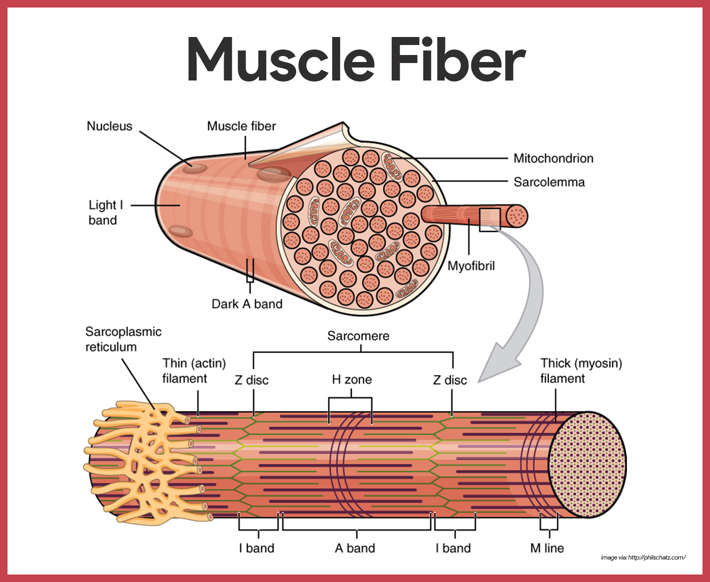
Skeletal Muscle Histology Slide Identification and Labeled Diagram ... Please try to find out these structures from the skeletal muscle slide labeled images. #1. Longitudinal section of skeletal muscle #2. Cross-section of skeletal muscle #3. Skeletal muscle fibers of the longitudinal section #3. The nucleus of skeletal muscle fibers in longitudinal and cross-section #4. Cross striations of skeletal muscles #5. Skeletal Muscle Fiber Labeled Muscle skeletal transverse labeled tissue section. Skeletal Muscle Fiber Labeled. SIU SOM Histology SSB we have 9 Pics about SIU SOM Histology SSB like Slide 34 - Skeletal Muscle - YouTube, Microscopic structure of skeletal muscle by Dr. S. N. Singh and also Microscopic structure of skeletal muscle by Dr. S. N. Singh. Here you go: Skeletal Muscle Fiber Structure and Function - Open Textbooks for Hong Kong Within each muscle fiber are myofibrils, long cylindrical structures that lie parallel to the muscle fiber. Myofibrils run the entire length of the muscle fiber. They attach to the plasma membrane, called the sarcolemma, at their ends, so that as myofibrils shorten, the entire muscle cell contracts ( Figure 16.18 ). Skeletal Muscle Fiber Labeling Quiz - PurposeGames.com This is an online quiz called Skeletal Muscle Fiber Labeling. There is a printable worksheet available for download here so you can take the quiz with pen and paper. Your Skills & Rank. Total Points. 0. Get started! Today's Rank--0. Today 's Points. One of us! Game Points. 15.
3,380 Muscle Cells Stock Photos, Pictures & Royalty-Free ...
Skeletal Muscle | Anatomy and Physiology I - Lumen Learning Skeletal Muscle Fibers. Because skeletal muscle cells are long and cylindrical, they are commonly referred to as muscle fibers. Skeletal muscle fibers can be quite large for human cells, with diameters up to 100 μm and lengths up to 30 cm (11.8 in) in the Sartorius of the upper leg. During early development, embryonic myoblasts, each with its ...
6 BAB II TINJAUAN PUSTAKA 2.1 Sel Sel adalah unit struktural ...
Chapter 9 Muscular Homework Flashcards | Quizlet Label the structures found within a skeletal muscle. ... The functional connection between a neuron and a skeletal muscle fiber is a. synapse.

Muscular System Objectives: A.Describe the purpose of the ...
Skeletal Muscle Fiber Labeling Flashcards | Quizlet mitochondrion myofibril what is this whole element? dark a band light i band nucleus purple dots on muscle fiber thin (actin) filament thick (myosin) filament z disc h zone m line elastic (titin) filament muscle fiber a single muscle cell can also be called ... myofibril there are many __________ in a single muscle fiber/cell. sarcomere
Sarcomere Model | Muscle Fiber Model | Skeletal Muscle ...
Skeletal Muscle - Anatomy & Physiology - University of Hawaiʻi Skeletal Muscle Fibers. Because skeletal muscle cells are long and cylindrical, they are commonly referred to as muscle fibers. Skeletal muscle fibers can be quite large for human cells, with diameters up to 100 μm and lengths up to 30 cm (11.8 in) in the Sartorius of the upper leg.During early development, embryonic myoblasts, each with its own nucleus, fuse with up to hundreds of other ...
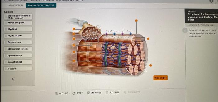
Solved Label structures associated with a neuromuscular ...
Correctly Label The Following Parts Of A Skeletal Muscle Fiber A skeletal muscle fiber is composed of a plasma membrane and a specialized smooth endoplasmic reticulum. It also contains sarcomeres and calcium ions. In addition to the plasma membrane, a skeletal muscle fiber has numerous myofibrils. During a contraction, the force is transmitted through the tendon to the bone, producing a skeletal movement.
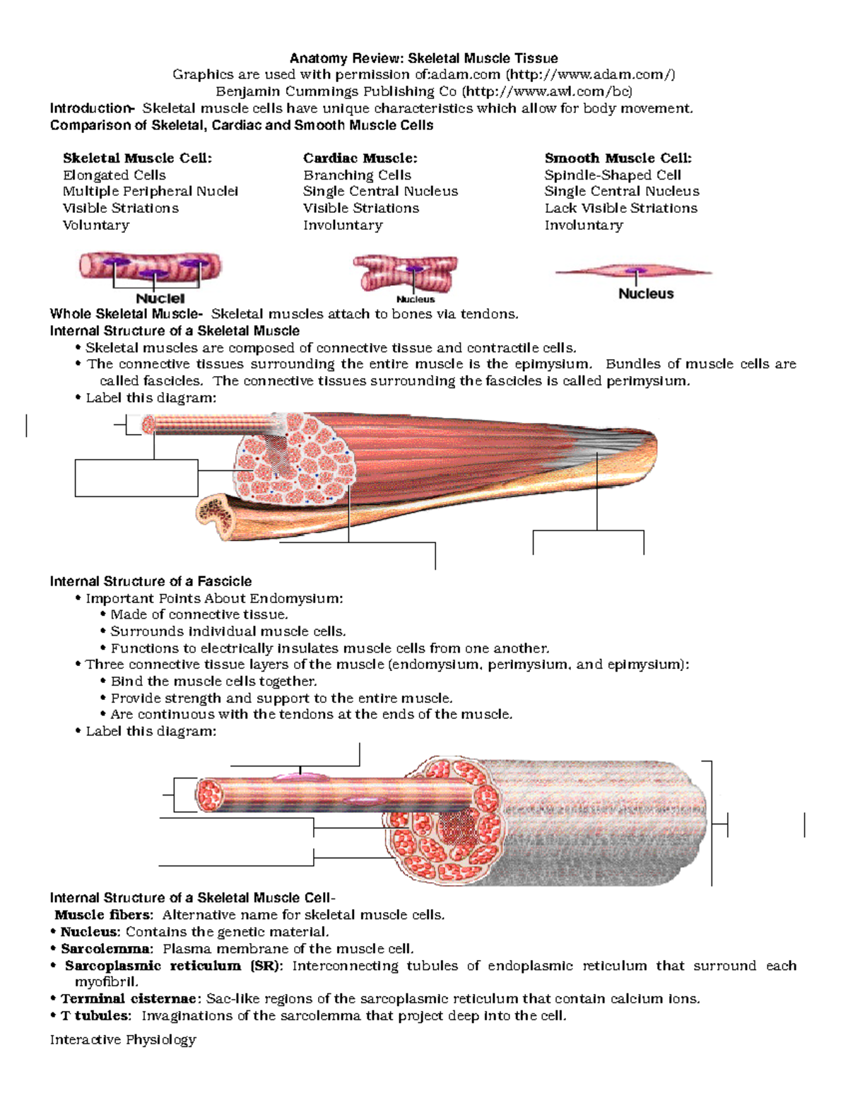
Anatomy Review: Skeletal Muscle Tissue
Muscle Fiber Anatomy Quiz - PurposeGames.com This is an online quiz called Muscle Fiber Anatomy. There is a printable worksheet available for download here so you can take the quiz with pen and paper. Your Skills & Rank. Total Points. 0. Get started! Today's Rank--0. Today 's Points. One of us! Game Points. 15. You need to get 100% to score the 15 points available.

Structure of a Skeletal Muscle Fiber Quiz
ZAKβ is activated by cellular compression and mediates ... Jul 28, 2022 · Skeletal muscle is a tissue where the role of this kinase is especially clear, and ZAKβ is strongly activated upon muscle fiber contraction. Besides the general implications for cell physiology, our discovery of ZAKβ function addresses important outstanding questions regarding how muscles sense and respond to the extent of mechanical load ...

The structure and internal organization of a skeletal muscle ...
Chapter 28 Reproductive System Flashcards | Quizlet Label the structures at the pointer tips in this view of the pelvic region. Match the region of the uterine tube with its description. Place the gene copy numbers below each cell to indicate how many copies of that gene are represented in each cell, and view meiosis from the standpoint of minimal gene copy numbers!

Types of Muscle Tissue and Fibers | Biology for Majors II
Skeletal Muscle Fiber Location and Arrangement | GetBodySmart Skeletal muscle fibers are located inside muscles, where they are organized into bundles called fascicles (= fasciculi). The epimysium is the connective tissue layer that covers the outer surface of the muscle. Surrounding and holding together each fascicle is a layer of connective tissue known as perimysium.
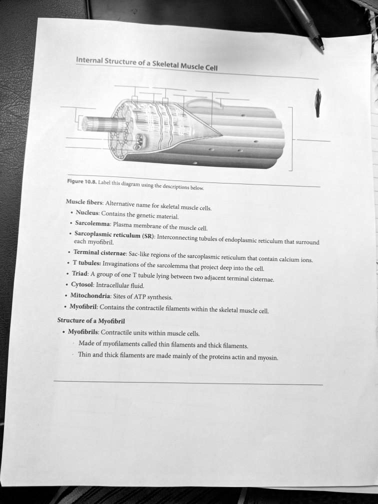
SOLVED: Internal Structure of a Skeletal Muscle Cell Figure ...
Phosphoproteomics of three exercise modalities identifies ... Jul 25, 2022 · One of these core phosphosites was S67 on the uncharacterized protein C18ORF25, which we validated as an AMPK substrate. Mice lacking C18ORF25 have reduced skeletal muscle fiber size, exercise capacity, and muscle contractile function, and this was associated with reduced phosphorylation of contractile and Ca 2+ handling proteins. Expression of ...

Muscle structure – muscle under the microscope — Science ...
Art-labeling Activity: The Structure of a Skeletal Muscle Fiber - Quizlet Start studying Art-labeling Activity: The Structure of a Skeletal Muscle Fiber. Learn vocabulary, terms, and more with flashcards, games, and other study tools.

Week 6: Muscle Physiology Flashcards | Quizlet
Skeletal Muscle Fiber - GetBodySmart A sarcomere is the smallest functional unit of skeletal muscle tissue, and each sarcomere has thick and thin filaments primarily composed of the proteins myosin and actin. The interaction of actin and myosin causes muscle contraction and therefore movement. Learn the physiology of skeletal muscles with the interactive tutorials and diagrams below.

Bellringer 12/9 Label #4 and #5. Building Skeletal Muscle… We ...
Label Skeletal Muscle Tissue Diagram | Quizlet bundles of myofilaments indise the muscle fiber. Muscle fiber one muscle cell. These are elongated cells with multiple peripheral nuclei. Myofilaments bundles of protein filaments inside the muscle fiber Actin this is the thin myofilament. Working together with myosin, it creates muscle contraction. Myosin this is the thick myofilament.
Answered: Art-labeling Activity: Structural… | bartleby
Muscle Fibers: Anatomy, Function, and More - Healthline Each one of your skeletal muscles is made up of hundreds to thousands of muscle fibers that are tightly wrapped together by connective tissue. Each muscle fiber contains smaller units made up of...
Fascicle Stock Illustrations – 240 Fascicle Stock ...
Structure of Skeletal Muscle | SEER Training Each skeletal muscle fiber is a single cylindrical muscle cell. An individual skeletal muscle may be made up of hundreds, or even thousands, of muscle fibers bundled together and wrapped in a connective tissue covering. Each muscle is surrounded by a connective tissue sheath called the epimysium.
Solved] Label all the parts of the follow diagrams. | Course Hero
Skeletal Muscle Fiber Definition and Anatomy - Study.com Structure of Skeletal Muscle Fiber Skeletal muscle fibers are composed of a bundle of thin filaments called myofibrils. Each myofibril is made up of small sections called sarcomeres. Sarcomeres are...
Untitled
Muscle Fiber Anatomy Strengthening lower trapezius. Muscle Fiber Anatomy. Structure of Muscle Fibers (IB Biology) - YouTube we have 9 Pics about Structure of Muscle Fibers (IB Biology) - YouTube like Skeletal Muscle Fiber, Cold Laser on Bicep Tendinitis Treatment and Shoulder Sprains and also MUSCULAR SYSTEM ANATOMY:Muscle fiber with neuromuscular junction model.

Study Guide Flashcards | Quizlet
11.2: Microscopic Anatomy of Skeletal Muscles - Biology LibreTexts Microscopic Anatomy of Skeletal Muscles. Skeletal muscle is found attached to bones. It consists of long multinucleate fibers. The fibers run the entire length of the muscle they come from and so are usually too long to have their ends visible when viewed under the microscope. The fibers are relatively wide and very long, but unbranched.
Frontiers | Tissue-Engineered Skeletal Muscle Models to Study ...
Solved Hel Label the structures of a skeletal muscle fiber ... - Chegg Science. Anatomy and Physiology. Anatomy and Physiology questions and answers. Hel Label the structures of a skeletal muscle fiber. 4 0.1 points eBook Sarcoplasmic reticulum Nucleus Myofibril Openings into T tubules Sarcolemma Mc Graw Hill < Prey 4 of 20 !!!
Skeletal Muscle: Longitudinal Section
Muscle Structure Labeling Quiz - PurposeGames.com This online quiz is called Muscle Structure Labeling. This online quiz is called Muscle Structure Labeling. English en. Login. Login Register Free Help; Start; Explore. Games; Playlists; Tournaments; Tags; The Wall; Badges; ... Skeletal Muscle Matching - Back of the Body 10p Image Quiz. Label Parts of the Skull - Lateral View 10p Image Quiz.

CH10HW Flashcards | Quizlet
Place the following steps in order of how a muscle contracts ...
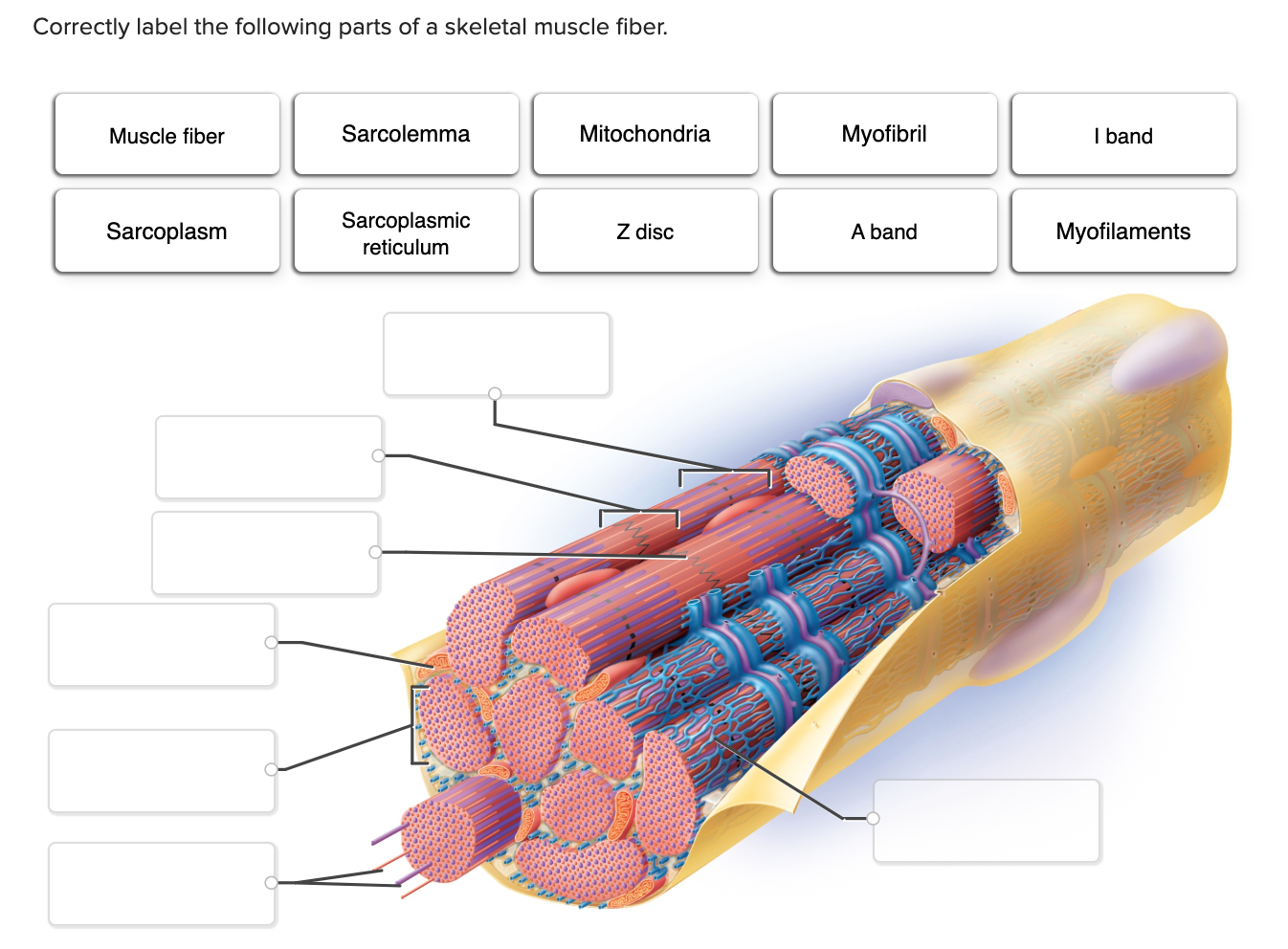
Solved Correctly label the following parts of a skeletal ...

10.2 Skeletal Muscle – Anatomy & Physiology

Structure of a skeletal muscle fiber Quiz
Frontiers | The Structure and Role of Intramuscular ...

Diagram of Skeletal Muscle
Skeletal muscle fiber hi-res stock photography and images - Alamy
Solved -ling Activity: Structure of a Skeletal Muscle Fiber ...
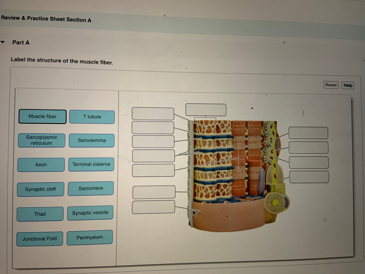
Answered: Label the structure of the muscle fiber | bartleby
Muscle Cell Without Labels, HD Png Download - kindpng
슬라이드 1
Anatomy Review Skeletal Muscle Tissue - Anatomy Review ...

Lesson Explainer: Structure of Muscles | Nagwa

Scheme of skeletal muscle and associated structures. (Left ...

Identify the structures of skeletal muscle. - Brainly.com
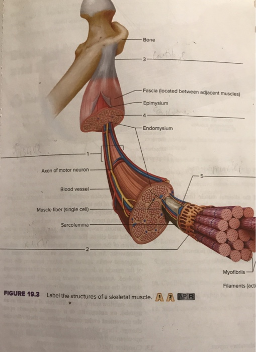
Solved Bone Fascia (located between adjacent muscles) | Chegg.com

Label structure of skeletal muscle Diagram | Quizlet
Muscular System Anatomy and Physiology - Nurseslabs
SISTEM OTOT MANUSIA
Post a Comment for "40 label the structures of a skeletal muscle fiber."