38 labelled diagram of compound microscope
Eye - Wikipedia Photoreception is phylogenetically very old, with various theories of phylogenesis. The common origin of all animal eyes is now widely accepted as fact.This is based upon the shared genetic features of all eyes; that is, all modern eyes, varied as they are, have their origins in a proto-eye believed to have evolved some 650-600 million years ago, and the PAX6 gene is considered a key factor in ... (a) Draw the labelled ray diagram for the formation of image ... Click here👆to get an answer to your question ️ (a) Draw the labelled ray diagram for the formation of image by a compound microscope. Derive an expression for its total magnification (or magnifying power), when the final image is formed at the near point.(b) Why both objective and eyepiece of a compound microscope must have short focal lengths?Draw a ray diagram showing the image ...
A compound microscope uses an objective lens of focal length ... (a) Draw the labelled ray diagram for the formation of image by a compound microscope. Derive an expression for its total magnification (or magnifying power), when the final image is formed at the near point. (b) Why both objective and eyepiece of a compound microscope must have short focal lengths?
Labelled diagram of compound microscope
Amazing 27 Things Under The Microscope With Diagrams May 13, 2022 · Observation under the compound microscope. Under a compound microscope, the differences between the sand particles become more apparent. It is visible that the shape, size color, and texture of individual particles vary within the sand collected from the same place. Some grains might appear smooth, while others appear irregular and sharp. Compound Microscope – Diagram (Parts labelled), Principle and ... Feb 03, 2022 · Image : Labeled Diagram of compound microscope parts. See: Labeled Diagram showing differences between compound and simple microscope parts Structural Components. The three structural components include. 1. Head. This is the upper part of the microscope that houses the optical parts. 2. Arm Inverted Microscope - Advantages, Disadvantages ... The inverted is offered with two models - routine and research. An upright microscope can be purchased as a smaller model for student use which influences price. History. A Dutch spectacle maker named Zaccharias Janssen invented the first microscope in about 1590. Merely a series of lens in a tube, it was the forerunner of today’s compound ...
Labelled diagram of compound microscope. Top 16 Techniques Used in Cell Biology (With Diagram) ADVERTISEMENTS: The following points highlight the top sixteen techniques used in cell biology. Some of the techniques are: 1. Immunofluorescence Microscopy 2. Ion-Exchange Chromatography 3. Affinity Chromatography 4. Partition and Adsorption Chromatography 5. Gel Filtration Chromatography 6. Radioactive Tracer Technique 7. Radioimmunoassay (RIA) 8. Enzyme Immunoassay 9. Spectroscopy and ... Inverted Microscope - Advantages, Disadvantages ... The inverted is offered with two models - routine and research. An upright microscope can be purchased as a smaller model for student use which influences price. History. A Dutch spectacle maker named Zaccharias Janssen invented the first microscope in about 1590. Merely a series of lens in a tube, it was the forerunner of today’s compound ... Compound Microscope – Diagram (Parts labelled), Principle and ... Feb 03, 2022 · Image : Labeled Diagram of compound microscope parts. See: Labeled Diagram showing differences between compound and simple microscope parts Structural Components. The three structural components include. 1. Head. This is the upper part of the microscope that houses the optical parts. 2. Arm Amazing 27 Things Under The Microscope With Diagrams May 13, 2022 · Observation under the compound microscope. Under a compound microscope, the differences between the sand particles become more apparent. It is visible that the shape, size color, and texture of individual particles vary within the sand collected from the same place. Some grains might appear smooth, while others appear irregular and sharp.
Microscope Parts Quiz

Simple Columnar Epithelium: Description: Single layer of elongated ...

Deaf Scientist Corner

Free Microscope Drawing, Download Free Microscope Drawing png images ...
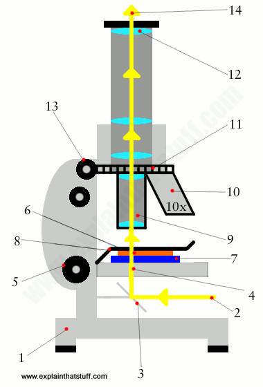
How does a microscope work? - Explain that Stuff

Labeling the parts of the microscope. (Blank diagram available for ...

Compound Light Microscope
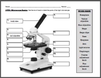
Quiz: Microscope Basics by Cynthia Zack | Teachers Pay Teachers

Mikroskop Nedir? Nasıl Kullanılır? | FenEhli.com
Post a Comment for "38 labelled diagram of compound microscope"